
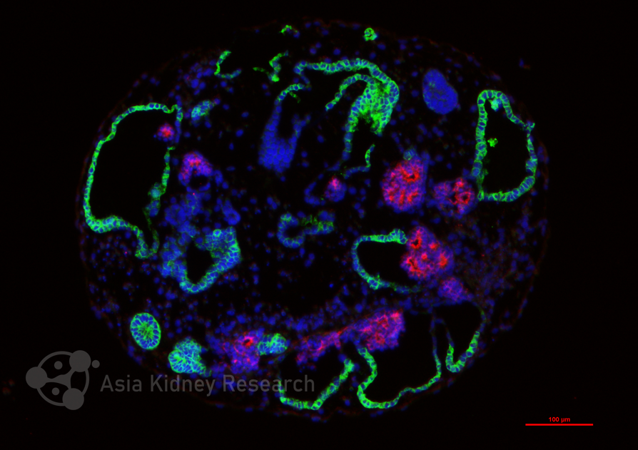
Figure: Kidnioid® ADPKD human kidney organoids from Asia Kidney Research.
The dilemma in drug development for autosomal dominant polycystic kidney disease (ADPKD) is not a lack of targets, but a lack of a validation system that can simultaneously answer "does it work" and "why does it work" in a human context. After tolvaptan, the pipeline appears prosperous—metabolism, inflammation, fibrosis, and gene therapy routes running in parallel—but very few truly reach clinical endpoints. Organoids are held in high hopes, but after sorting through the existing evidence, a more sober judgment emerges: the value of organoids lies not in "replacing animals," but in "blocking compounds that should not move forward at the right position, and letting through true drugs that were mistakenly killed by old models" [1-3]. The premise for this judgment to hold is to clarify what each model can answer and what it cannot answer.
I. Where ADPKD Drug Development Stands: Walking on One Leg, Testing with Several Others
Tolvaptan is the only approved disease-modifying drug, but it only covers rapid progressors, and its aquaresis and hepatotoxicity limit widespread use [4,5]. Later entrants in the pipeline fall roughly into four categories, each corresponding to different validation needs.
The metabolic route is currently the most crowded track. The Phase III trial of metformin (IMPEDE-PKD) is ongoing, and sodium-glucose cotransporter 2 (SGLT2) inhibitors, glucagon-like peptide-1 (GLP-1) receptor agonists, and bempedoic acid are all in early clinical or preclinical stages [5-7]. The logic of this route is: cyst epithelia depend on glycolysis and have impaired mitochondrial function, so correcting metabolic status may delay progression [8-10]. Readout endpoints for such drugs, besides cyst size, include adenosine triphosphate (ATP) levels, oxygen consumption rate/extracellular acidification rate (OCR/ECAR), lipid content, and autophagic flux [9,11,12].
The cyclic adenosine monophosphate (cAMP) axis is still extending. Beyond tolvaptan, long-form phosphodiesterase 4 (PDE4) activators attempt to precisely lower cAMP within cyst microdomains, theoretically avoiding the systemic aquaresis of vasopressin V2 receptor (V2R) antagonists [13,14]. The validation premise for this route is that the model must have functional cAMP-protein kinase A (PKA) signaling and preferably express the V2 receptor (AVPR2)—otherwise even "whether lowering cAMP is sufficient" cannot be measured accurately [15,16].
Anti-inflammatory/anti-fibrotic is theoretically a "combination punch" direction. Ligand-receptor pairs such as macrophage migration inhibitory factor (MIF), secreted phosphoprotein 1 (SPP1), and tumor necrosis factor-like weak inducer of apoptosis (TWEAK) have been repeatedly identified in human ADPKD spatial transcriptomics, suggesting active dialogue between cyst epithelia and fibroblasts and macrophages [17-19]. But the validation dilemma for such targets is: pure in vitro organoids contain no fibroblasts or macrophages. An anti-MIF antibody may be completely "ineffective" in nephron organoids (NKO)—not because the target is wrong, but because the model lacks the cells to receive the signal [20,21].
Gene therapy is the most "mechanism-based" direction, and also the hardest to validate. Anti-microRNA-17 (anti-miR-17) oligonucleotide (farabursen) has entered Phase III, the polycystin-1 (PC1) folding corrector (VX-407) targeting PKD1 missense mutations has entered Phase II, and base editing has been shown in organoids to reverse cystic phenotypes [5,22-24]. The core validation question for this route is: whether the corrected protein truly restores function, not merely "is expressed." This requires a model that can distinguish "protein present" from "protein working on the cilium" [23,25].
The pipeline is widening, but depth is narrowing. ADPKD is not a large market like oncology, but it has an advantage rare diseases do not: clear targets, concentrated pathways, and enrichable patient cohorts [5,26]. This has attracted many "old drug repurposing" attempts (metformin, SGLT2 inhibitors, GLP-1 receptor agonists), as well as high-risk, high-reward directions like gene therapy. Judging from paper counts and clinical trial registrations, the field's heat continues to rise. But the heat is unevenly distributed—what truly reaches Phase III remains the cAMP axis and the metabolic axis; anti-inflammatory/anti-fibrotic and protein homeostasis directions, though conceptually valid, remain early because of the lack of models capable of validating them [2,5].
II. The Real Capability Boundaries of Organoid Models: Four Models, Four "Answer Only One Question"
Existing organoid models can be divided into four tiers according to capability boundaries. Each tier has clear "can" and "cannot" limits, and confusing the tiers is the most common mistake in drug development [1,2,27].
NKO (nephron organoids) are a "broad screening machine," not a "mechanism-based judge." Their advantages are clear: high throughput, rapid cyst formation, and controllable cost [28-30]. Tran et al. used mini-NKOs (about 1,500 cells per well, 1–2 nephron-like structures per organoid, about 4,000 organoids per 96-well plate) to screen 247 protein kinase inhibitors and identified QNZ, with half-maximal inhibitory concentration (IC50) values of 6.6 nM and 4.5 nM against PKD1⁻/⁻ and PKD2⁻/⁻ organoids, respectively [28]. Another study achieved scalable production using ureteric bud (UB) organoid suspension culture—dissociating about 50 nephron organoids to produce up to 2,000 UB organoids within 7 days—and on this basis screened 305 nutrient-like molecules, identifying the Toll-like receptor 4 (TLR4) antagonist M1; pathway validation was then performed with the known TLR4 inhibitor TAK-242, observing dose-dependent cyst reduction by TAK-242, cyst enlargement by lipopolysaccharide (LPS), and reversal of LPS effects by TAK-242 [31].
Two things need to be distinguished here: M1 is a new hit from library screening, while TAK-242 is a known tool compound validating the TLR4 pathway; their roles differ [31]. The readout for NKO/UB organoids is cyst formation; inhibition observed at this level reflects the compound's effect on the cystic phenotype. NKOs do not express AVPR2, and tolvaptan produces no inhibitory effect in such models [15,28,31]; the same compound shows cyst inhibition in CD organoids [15]. The difference lies in whether the model expresses AVPR2.
Collecting duct (CD) organoids are the "gatekeepers of mechanism-based validation," but their throughput does not allow them to do broad screening. Among current evidence, CD organoids are one of the few models that produce an inhibitory response to tolvaptan in vitro, because they express AVPR2 [15]. Mae et al. expanded human induced pluripotent stem cell (hiPSC)-derived UB tip cells (UBTC) long-term and differentiated them into CD organoids containing cortical collecting duct principal cells; PKD1⁻/⁻ organoids formed cysts spontaneously, arginine vasopressin (AVP) enhanced cystic expansion, and tolvaptan (5 μM) significantly inhibited AVP-induced cyst enlargement [15]. Small-scale compound testing on the same platform also showed that retinoic acid receptor (RAR) agonists (TTNPB, all-trans retinoic acid [ATRA]) inhibited cyst expansion, and ATRA also improved kidney weight ratio and blood urea nitrogen (BUN) in Pkd1flox/flox;Ksp-Cre mice [15]. It should be noted that the human advancement of RAR agonists (tamibarotene Phase IIa) was the result of subsequent clinical research and should not be phrased as a single-line causal claim that it was "directly screened out from this CD organoid platform and went to clinic" [15,32]. CD organoids have long preparation cycles and lower throughput than mini-NKOs; their reasonable position is secondary screening and mechanistic confirmation, not replacement of broad screening [1,15].
Patient-derived multi-lineage adult renal organoids (MARO) and tubuloids answer "genotype–drug efficacy" questions, not "throughput" questions. MARO are derived directly from patient cyst wall tissue and retain native mutations and multiple lineages (proximal tubule [PT], thick ascending limb [TAL], collecting duct [CD]) [20]. Guo et al. used patient-derived MARO to observe that tolvaptan was more effective in PKD1 mutants and less effective in PKD2, while Rho inhibitors (ML141, EHT 1864, CCG-1423) were effective across genotypes [20]. If primary screening compounds need to distinguish "which patients are more likely to respond," MARO can provide this information. But their throughput determines that they are suitable for validation, not discovery. It should be qualified that the above genotype differences come from specific patient lines and experimental conditions in that study and cannot yet be generalized as a universal rule for all PKD1/PKD2 mutations [20].
Human kidney progenitor assembloid (hKPA) in vivo transplantation is the "last gate before clinical translation," not a "drug screening tool." Huang et al. transplanted PKD2⁻/⁻ hKPA under the renal capsule of NOD/SCID/IL-2Rγ⁻/⁻ (NSG) mice; after 2 months, grafts developed cysts (cyst fluid containing creatinine and urea), myofibroblast layers, CD45⁺/F4/80⁺ macrophage infiltration, and human-derived vessels, with cyst epithelial cilia facing the cyst lumen, consistent with patients [21]. In contrast, cyst epithelia in pure in vitro NKOs often show apical-out cilia, an in vitro polarity artifact [30,33]. A compound claiming to "both shrink cysts and resist fibrosis" may perform well in NKOs but fail or show toxicity in hKPA due to immune microenvironment involvement [21]. The position of hKPA is not to replace NKO screening, but to make a final "is it worth continuing" judgment in a context closer to patients before compounds enter clinical development. Its limitations are equally clear: low throughput, high cost, dependence on animal transplantation, unsuitable as a routine screening platform [21].
III. Mechanisms Covered by Models: A "Blank Map" That Needs to Be Taken Seriously
If the known mechanisms of ADPKD are listed and compared with model capabilities, some structural gaps emerge [1,2,27].
The cAMP/AVPR2 axis is best covered, but also most "crowded." NKO, CD organoids, tubuloids, and MARO can all measure cAMP-related readouts [15,20,34,35]. But this also means that for new drugs along this axis, the "incremental information" from organoids is limited—tolvaptan has already proven this route, and later PDE4 activators or novel V2R antagonists can be relatively validated with existing models [13,14].
The Rho/planar cell polarity (PCP)/cilia-dependent cyst activation (CDCA) axis is currently systematically validated mainly in MARO. Guo et al. used intraflagellar transport 88 (IFT88) knockout in patient MARO to demonstrate cilia-dependent cyst activation and positioned Rho/PCP as downstream effectors [20]. But whether this holds similarly in tubuloids, CD organoids, and hKPA, current evidence does not answer. This is a clear gap: if Rho inhibitors are to move toward clinical use, the universality of the CDCA mechanism needs to be confirmed across multiple models.
The TLR4/nuclear factor kappa-B (NF-κB) axis is currently validated mainly in UB organoids. Vanichapol et al.'s TAK-242 data support the involvement of the TLR4 pathway in cyst size regulation, but the system is PKD2-knockout, suspension-cultured UB organoids [31]. Whether TLR4 similarly drives cysts in CD organoids, MARO, and hKPA remains unclear. Natural ligands of TLR4 partly come from injury and inflammation, and pure epithelial organoids may underestimate or overestimate its role; therefore, if TLR4 inhibitors are to advance, they need to be revalidated in models with an immune microenvironment [21,31].
Metabolic reprogramming has cross-model evidence, but "metabolic endpoints" are ignored in most drug screens. Huang et al. measured elevated OCR and ECAR in Pkd2⁻/⁻ induced nephron progenitor cell (iNPC)-derived organoids (no difference in undifferentiated NPCs) [12]; Mae et al. measured changes in fatty acid oxidation, cholesterol/bile acid metabolism, and ATP levels in CD organoids [15]; Wang et al. measured decreased mitochondrial DNA copy number in patient iPSC-derived organoids (no difference in iPSCs themselves) [23]. In preclinical studies of metabolic intervention in ADPKD, efficacy assessment often focuses on both cyst burden and metabolic indicators—ATP production, glycolytic flux, mitochondrial respiration, and autophagic status [9,11,12].
Fibrosis/immune/vascular currently can only be covered by the hKPA in vivo model. This is a hard boundary, with no in vitro alternative [21]. Any ADPKD candidate drug claiming "anti-fibrotic" effects, if validated only in in vitro organoids, has an incomplete preclinical evidence chain [21,30].
Protein homeostasis (transmembrane emp24 protein transport domain-containing 9 [TMED9]; ALG8; unfolded protein response [UPR]) is almost untouched in ADPKD organoids. The discovery of TMED9/BRD4780 came from MUC1 kidney disease in autosomal dominant tubulointerstitial kidney disease (ADTKD) [25], and the ALG8 metabolic threshold model remains at the review level [36]. The path "PKD1 missense mutation → protein folding defect → endoplasmic reticulum stress (ER stress)" has not been systematically validated in ADPKD organoids [25,36]. If folding correctors like VX-407 are to advance, an organoid system that can distinguish "protein expression" from "protein reaching the cilium and working" is needed—currently no such evidence exists [23,25].
IV. How to Use Them: A Sequential, Trade-off "Funnel," Not a Bunch of Parallel Options
Drug development resources are limited, and the preparation cost and time of organoids do not allow "trying every model once." An effective strategy is to allocate models by stage, not to choose models by preference [1,2].
Stage 1: Discovery. Use mini-NKOs or UB organoid suspension culture to screen large libraries [28,31]. Positive compounds at this step only mean "can inhibit cysts," not "mechanism-based." Tolvaptan is usually negative at this step and should not be used to reject compounds [15,28]. Both QNZ and M1 came out of this stage, but their subsequent validation paths differ—QNZ needs CD organoids to confirm whether NF-κB similarly drives cysts in collecting duct epithelium; M1/TAK-242 needs MARO or hKPA to confirm the role of TLR4 in the immune microenvironment [15,20,21,28,31].
Stage 2: Mechanism-based secondary screening. Put Stage 1 candidates into CD organoids (if the target is cAMP/AVPR2) or patient MARO (if the target is Rho/PCP or genotype distinction is needed) [15,20]. The purpose of this step is to eliminate compounds that "work only in the fetal-like epithelium of NKOs but fail in mature collecting ducts or patient mutation backgrounds." RAR agonists validated in CD organoids for cyst inhibition are an example of the value of such secondary screening; but their subsequent clinical advancement also involves independent human trials and cannot be simplified as "organoids sent directly to clinic" [15,32].
Stage 3: In vivo confirmation. For compounds entering preclinical candidacy, assess overall benefit and safety in hKPA transplantation or animal models [21,37,38]. The key at this step is not "whether cysts got smaller," but "whether cyst shrinkage is accompanied by improved renal function, whether fibrosis is inhibited, and whether toxicities invisible in vitro appear." The value of hKPA lies in providing the immune and vascular context missing in vitro, giving claims like "anti-fibrotic" and "anti-inflammatory" a place to be tested [21].
Parallel track: Metabolic and protein homeostasis drugs need different readout endpoints. Metabolic drugs measure OCR/ECAR/ATP in CD organoids or NKOs while retaining cyst size as a parallel readout [9,11,12,15]; protein homeostasis drugs need patient iPSC organoids with PKD1 missense mutations to measure whether PC1 reaches the cilium and whether ER stress is relieved [23,25,36]. Screening with cyst size as the primary endpoint looks at whether cysts got smaller; metabolic drugs and protein homeostasis drugs need to answer whether the metabolic pathway has been corrected and whether the protein has reached the cilium.
Conclusion
The most honest conclusion from current evidence is: ADPKD organoids are already good enough to help us make decisions, but far from good enough to make decisions for us [1-3]. NKOs help us screen broadly, CD organoids help us confirm mechanism, MARO help us distinguish patients, and hKPA help us gate preclinical development. Each model answers only one question, and failures in drug development often occur when "a model that answers question A is used to answer question B" [1,2,27].
After tolvaptan, the ADPKD pipeline is widening, but true breakthroughs will not come from "yet another compound screened out by NKOs," but from putting the right compounds into the right models and making the right trade-offs at the right stage [2,5]. Organoids are not the answer; they are the tool that lets us ask the right questions.
References
Dixon EE, Woodward OM. Three-dimensional in vitro models answer the right questions in ADPKD cystogenesis. Am J Physiol Renal Physiol. 2018;315(2):F332-F335.
Afrin H, Qamar U, Robichaud JH, et al. Kidney organoid models of polycystic kidney disease: challenges and future directions. Am J Physiol Renal Physiol. 2025;329(1):F143-F159.
Scarlat A, Tomasoni S, Trionfini P. Autosomal dominant polycystic kidney disease: from pathogenesis to organoid disease models. Biomedicines. 2025;13(7):1766.
Torres VE, Chapman AB, Devuyst O, et al. Tolvaptan in patients with autosomal dominant polycystic kidney disease. N Engl J Med. 2012;367(25):2407-2418.
Chen CY, Hadla M, Khambati I, et al. Emerging therapies in autosomal dominant polycystic kidney disease. Kidney360. 2026;7:1662-1676.
Perrone RD, Abebe KZ, Watnick TJ, et al. Primary results of the randomized trial of metformin administration in polycystic kidney disease (TAME PKD). Kidney Int. 2021;100(3):684-696.
Capelli I, Lerario S, Ciurli F, et al. Investigational agents for autosomal dominant polycystic kidney disease: preclinical and early phase study insights. Expert Opin Investig Drugs. 2024;33(5):469-484.
Rowe I, Chiaravalli M, Mannella V, et al. Defective glucose metabolism in polycystic kidney disease identifies a new therapeutic strategy. Nat Med. 2013;19(4):488-493.
Lian X, Wu X, Li Z, et al. The combination of metformin and 2-deoxyglucose significantly inhibits cyst formation in miniature pigs with polycystic kidney disease. Br J Pharmacol. 2019;176(5):711-724.
Padovano V, Podrini C, Boletta A, Caplan MJ. Metabolism and mitochondria in polycystic kidney disease research and therapy. Nat Rev Nephrol. 2018;14(11):678-687.
Liu M, Zhang C, Gong X, et al. Kidney organoid models reveal cilium-autophagy metabolic axis as a therapeutic target for PKD both in vitro and in vivo. Cell Stem Cell. 2024;31(1):52-70.e8.
Huang B, Zeng Z, Kim S, et al. Long-term expandable mouse and human-induced nephron progenitor cells enable kidney organoid maturation and modeling of plasticity and disease. Cell Stem Cell. 2024;31(6):921-939.e17.
Omar F, Findlay JE, Carfray G, et al. Small-molecule allosteric activators of PDE4 long form cyclic AMP phosphodiesterases. Proc Natl Acad Sci U S A. 2019;116(27):13320-13329.
Zhou X, Torres VE. Emerging therapies for autosomal dominant polycystic kidney disease with a focus on cAMP signaling. Front Mol Biosci. 2022;9:981963.
Mae SI, Hattanda F, Morita H, et al. Human iPSC-derived renal collecting duct organoid model cystogenesis in ADPKD. Cell Rep. 2023;42(12):113431.
Kuraoka S, Tanigawa S, Taguchi A, et al. PKD1-dependent renal cystogenesis in human induced pluripotent stem cell-derived ureteric bud/collecting duct organoids. J Am Soc Nephrol. 2020;31(10):2355-2371.
Khan MI, Robichaud JH, Afrin H, et al. The polycystic kidney disease cyst transcriptome defined by integrating spatial, single nuclear, and bulk transcriptomics. Kidney360. 2026; doi:10.34067/KID.0000001371.
Chen L, Zhou X, Fan LX, et al. Macrophage migration inhibitory factor promotes cyst growth in polycystic kidney disease. J Clin Invest. 2015;125(6):2399-2412.
Cordido A, Nuñez-González L, Lamas-González O, et al. TWEAK signaling pathway blockade slows cyst growth and disease progression in autosomal dominant polycystic kidney disease. J Am Soc Nephrol. 2021;32(8):1913-1932.
Guo L, Fan Y, Sun H, et al. Patient-derived kidney organoids recapitulate ADPKD and facilitate the identification of Rho pathway inhibitors as candidate therapeutics. Cell Rep Med. 2026;7(4):102720.
Huang B, Medina P, He J, et al. Spatially patterned kidney assembloids recapitulate progenitor self-assembly and enable high-fidelity in vivo disease modeling. Cell Stem Cell. 2025;32(10):1614-1633.e13.
Lakhia R, Ramalingam H, Chang CM, et al. PKD1 and PKD2 mRNA cis-inhibition drives polycystic kidney disease progression. Nat Commun. 2022;13(1):4765.
Wang J, Qiu Y, Zhang L, et al. Adenine base editor corrected ADPKD point mutations in hiPSCs and kidney organoids. Adv Biotechnol. 2024;2:20.
Vishy CE, Thomas C, Vincent T, et al. Genetics of cystogenesis in base-edited human organoids reveal therapeutic strategies for polycystic kidney disease. Cell Stem Cell. 2024;31(4):537-553.e5.
Dvela-Levitt M, Shaw JL, Greka A. A rare kidney disease to cure them all? Towards mechanism-based therapies for proteinopathies. Trends Mol Med. 2021;27(4):355-369.
Bergmann C, Guay-Woodford LM, Harris PC, et al. Polycystic kidney disease. Nat Rev Dis Primers. 2018;4(1):50.
Kuzinska MZ, Lin SYY, Kiambt V, et al. Ciliopathy organoid models: a comprehensive review. Am J Physiol Cell Physiol. 2024;327(6):C1590-C1616.
Tran T, Song CJ, Nguyen T, et al. A scalable organoid model of human autosomal dominant polycystic kidney disease for disease mechanism and drug discovery. Cell Stem Cell. 2022;29(7):1083-1101.e7.
Freedman BS, Brooks CR, Lam AQ, et al. Modelling kidney disease with CRISPR-mutant kidney organoids derived from human pluripotent epiblast spheroids. Nat Commun. 2015;6:8715.
Cruz NM, Song X, Czerniecki SM, et al. Organoid cystogenesis reveals a critical role of microenvironment in human polycystic kidney disease. Nat Mater. 2017;16(11):1112-1119.
Vanichapol T, Wangford R, Mishra S, et al. Large-scale organoid-derived cyst cultures as a drug discovery platform for polycystic kidney disease. Kidney Int. 2026; doi:10.1016/j.kint.2026.07.034.
Shimizu T, Mae SI, Araoka T, et al. A novel ADPKD model using kidney organoids derived from disease-specific human iPSCs. Biochem Biophys Res Commun. 2020;529(4):1186-1194.
Dixon EE, Bradford STJ, Humphreys BD. Mini kidney organoids deliver maximal drug screening impact. Cell Stem Cell. 2022;29(7):1011-1012.
Xu Y, Kuppe C, Perales-Patón J, et al. Adult human kidney organoids originate from CD24+ cells and represent an advanced model for adult polycystic kidney disease. Nat Genet. 2022;54(11):1690-1701.
Rohe J, Kariat Ashraf AP, Kehl M, et al. Modelling kidney cystogenesis using human kidney tubuloid cultures. BMC Biol. 2026;24:26.
Wang X, Huang Z, Zhang W. ALG8-driven metabolic reprogramming in polycystic kidney disease: a systematic synthesis of evidence linking glycosylation defects to metabolic signaling. IUBMB Life. 2026;78(3):e70001.
Sieben CJ, Harris PC. Experimental models of polycystic kidney disease: applications and therapeutic testing. Kidney360. 2023;4(8):1155-1173.
Ramos AM, Fernandez-Fernandez B, Perez-Gomez MV, et al. Design and optimization strategies for the development of new drugs that treat chronic kidney disease. Expert Opin Drug Discov. 2020;15(1):101-115.